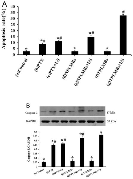
Figure 4.
Apoptosis efficiency in A2780/DDP cells with different treatments. (A) The percentage of apoptosis cells was determined by flow cytometry 24 h after transfection. Data are represented as mean ±SD (n=3). Apoptosis efficiency of the PLMBs +US groups and TPLMBs +US groups are significantly higher than those of the other groups (P<0.05). Apoptosis efficiency of TPLMBs +US group is higher than PLM+US group (P<0.05). Compared with group (g), P<0.05; Compared with group (a), #P<0.05. (B) Western blot analysis of the expression of Caspase-3 protein in A2780/DDP cells after different treatments. Caspase-3 protein expression was analyzed by Western blot 24 h after transfection. Cells treated with TPLMBs +US groups showed more prominent bands than other treatment groups (p<0.05). GAPDH was used as an internal reference. Compared with group (g), *p<0.05; Compared with group (a), #p<0.05.

