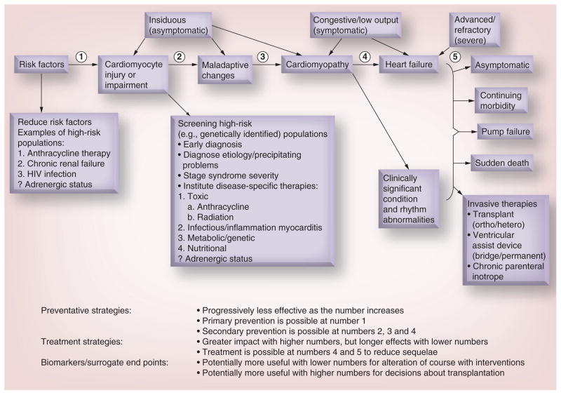
Figure 1. Stages in the course of pediatric ventricular dysfunction.
Review of the stages in the course of pediatric ventricular dysfunction that can be followed by echocardiographic measurements of left ventricular structure and function in conjunction with cardiac biomarkers that have been validated as surrogates for clinically significant cardiac end points. The identification of risk factors and high-risk populations for ventricular dysfunction are highlighted where their use may lead to preventive or early therapeutic strategies, while the determination of etiology may lead to etiology-specific therapies. Points (1–5) indicate stage-related points of intervention for preventive and therapeutic strategies, and where biomarkers and surrogate markers may be used. ? indicates that the role of adrenergic status is unknown.
Reproduced with permission from [21].

