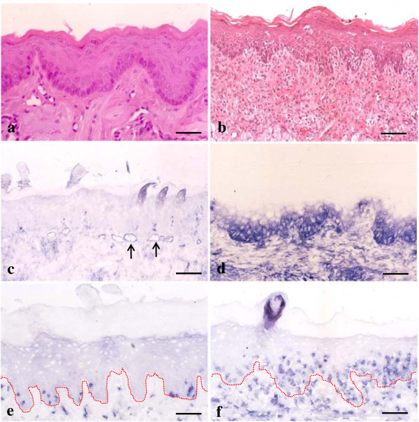
Figure 1.
Immunohistochemical expression of intercellular adhesion molecule-1 (ICAM-1) and CD8+ in graft-versus-host disease (GVHD)-related oral mucosa. a and b: No obvious changes are observed in hematoxylin and eosin (H&E)-stained control tongue sections (a). In tongue tissue sections from GVHD rats, epithelial destruction is observed (b). c and d: In tongue specimens from control rats, ICAM-1 is expressed only in the vascular slits (arrows) (c). Epithelial ICAM-1 expression is observed in the basal to spinous layers of the oral mucosa from GVHD rats (d). e and f: Only a few CD8+ cells are found in the control (e). In the GVHD rats, increased numbers of CD8+ cells are observed in both lamina propria and surface epithelium (f). The dotted line shows a junction between the surface epithelium and lamina propria of the tongue. Bar = 100 μm.

