Figure 2.

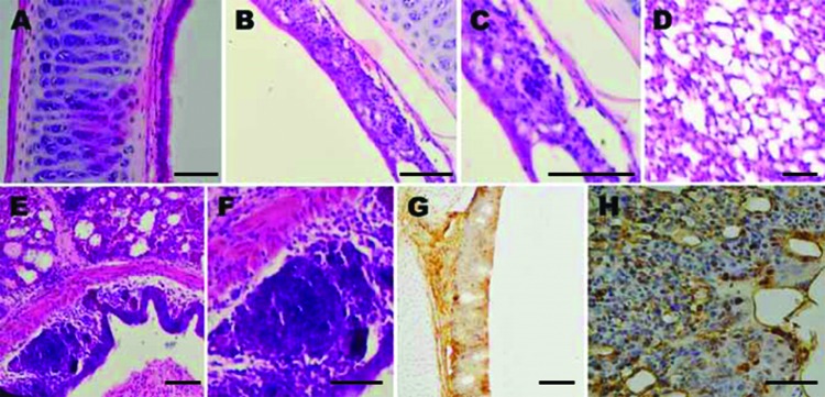
Histological appearance and immunohistochemical staining of respiratory tract samples collected from chickens before and after inoculation with avian metapneumovirus (aMPV) subgroup C, China. A) Trachea section from an uninoculated chicken shows intact ciliated epithelium. B) At 5 days’ postinoculation, loss of cilia, architectural disruption, and infiltration of inflammatory cells were seen in most of the epithelium and submucosa of inoculated chickens. C) Same lymphoid cell infiltration in trachea as in panel B, showing large numbers of lymphocytes in the epithelium. D) Lung section from an uninoculated chicken shows no significant inflammation. E) At 5 days’ postinoculation, inflammatory infiltration including lymphocytes, as well as scattered macrophages and heterophils were seen in most of the lungs of inoculated chickens. F) Same inflammatory infiltration in lung as in panel E, showing large numbers of lymphocytes in the bronchial submucosa of lung. G) Trachea tissue of aMPV-inoculated chicken shows many positive cells for aMPV antigen. H) Lung tissue of aMPV-inoculated chicken shows many positive cells for aMPV antigen. Scale bars indicate 80 μm.
