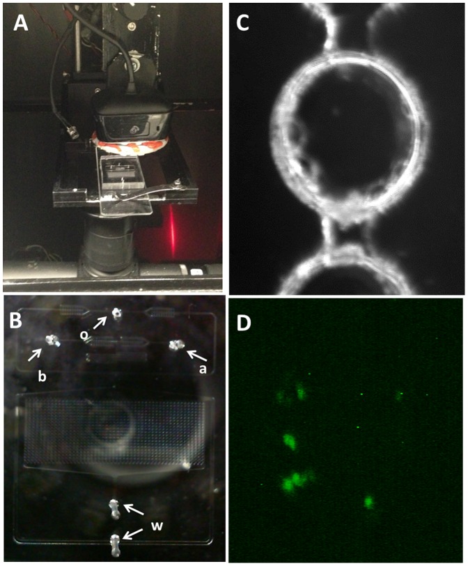
Figure 4. ScanDrop: E. coli detection.
A) Microfluidic chip operating inside the imaging system. B) Droplet array as viewed from the top camera. Arrows indicate tubing/chip connection locations as follows: o- oil (inlet); b- beads conjugated with E. coli (inlet); a- fluorescently labeled antibody (inlet); w- waste (outlets). C) Droplet image as seen from the top camera with white LED illumination. D) Antibody green fluorescence indicates the presence of RFP-expressing E. coli (positive control).

