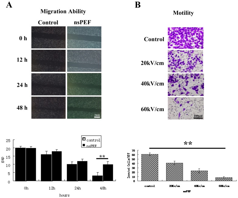
Figure 3. The motility and migration capability inhibition is dose-dependent in vitro.
At 1×10⁴ cells/well were seeded in the the upper chamber in the serum-free media. The lower chamber were filled with media supplemented with serum. The chambers were incubated at 37°C with 5% CO2. The cells with migratory capability moved through the pores toward the serum below. After 24 hours, migrated cells moved to the lower well were stained with crystal blue and photographed under a phase-contrast microscope in five randomly chosen fields (100×magnification). The experiments were performed in triplicate wells and performed three times. The pulsed cells were also perform wound assay to reflect tumor cell migration capability with scratches. After 12, 24 and 48 hours, the scratch area was photographed and the wound distance between edges was measured and averaged from 5 points per 1 wound area. Experiments were repeated three times. The nsPEFs treated cells showed decreased migration capability in a dose dependant manner (Figure 3A HepG2 under 40 kV/cm). Quantification analysis indicates reduced motility capability in a dose-dependent manner (Figure 3B HepG2 under 40 kV/cm). Similar results were also observed in SMMC7721, HCCLM3 and Hep1-6 cells (data not shown).

