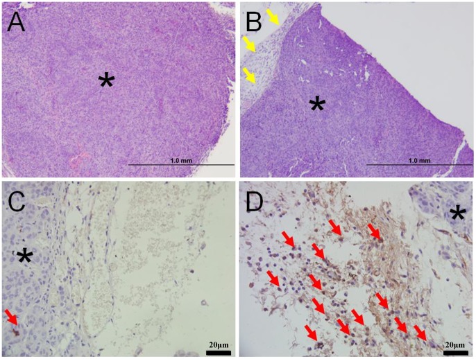
Figure 5. Macrophage infiltration contributes to the tumor ablation in vivo.
The tumors were dissected with the attached tissue to study the macrophages infiltration. The star indicate the tumor cells. H&E stain showed the one day after nsPEFs treatment, tumor in T100 group shrank dramatically (figure 5B) compared with S300 (Figure 5A). Immunolabelling of macrophages in formalin-fixed, paraffin-embedded tissue, using the ABC method and haematoxylin counter stain (figure 5A and 5B). Tumor nodule in T100 group (figure 5B) was surrounded by a remarkable infiltration of inflammatory cells (The yellow arrow) compared with S300 group (figure 5A) which demonstrated few inflammatory cells. MAC387 antibody stained a moderate to high number of macrophages in the periphery of tumor from T100 group (figure 5D). Very few MAC387 positive cells (The red arrow) was seen in the edge of tumor from S300 group (figure 5C); The high density of MAC387 positive cells (The red arrow) found in the leading edge of tumor and especially within perivascular areas reflect host defensive macrophages recruited into the tumor vicinity. Beside the typical macrophage staining in figure 5, the macrophage infiltration in 28 mice were also detected. There was no macrophage infiltration in 7 mice of CT (0/7), mild macrophage infiltration in 4 mice of S300 (4/7), macrophage infiltration in T100 (intense in 6 mice, 6/8, and mild in 2 mice, 2/8).

