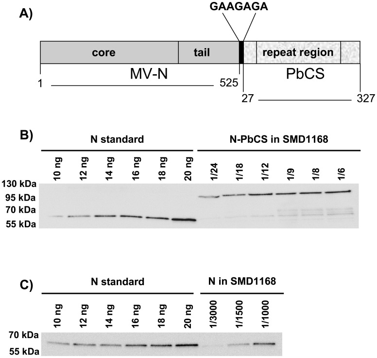
Figure 2. Expression of N-PbCS in P. pastoris.
(A) Schematic representation of N-PbCS fusion protein. MV-N (dark grey) is composed of a core domain in N-terminal and a unstructured tail domain in C-terminal [69]. The GAAGAGA linker is in black. PbCS (light grey) corresponds the central repeat region flanked by major portions of the N-terminal and C-terminal domains of the protein [39]. Amino acids numbering are given according to N from the MV Schwarz vaccine strain and PbCS from the Pb ANKA strain. For sequence details, see Figure S1. (B) Quantitative western blot analysis of SMD1168 expressing N-PbCS or (C) N. In (B) and (C), yeast lysates were diluted as indicated, the MV-N protein was used as a standard with increasing concentrations and western blots were probed with an anti-N antibody.

