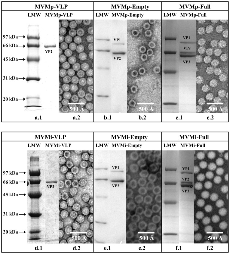
Figure 1. Virus purification.
Coomassie stained SDS-PAGE for MVMp-VLP (a.1), MVMp-Empty (b.1), MVMp-Full (c.1), MVMi-VLP (d.1), MVMi-Empty (e.1), MVMi-Full (f.1). The positions of low-molecular-weight (LMW) standards (in kDa; Bio-Rad, Hercules, CA, USA) are indicated on the left-hand side. Negatively stained electron micrographs are shown in (a.2), (b.2), (c.2), (d.2), (e.2), and (f.2) respectively. Images in (b.2) and (c.2) were collected at 60,000X magnification while images in (a.2), (d.2), (e.2), and (f.2) were collected at 100,000X magnification.

