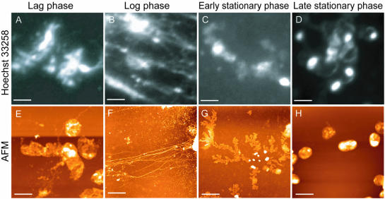
Figure 1.
Overall structure of the nucleoid. Escherichia coli strain W3110 was grown in LB medium. The cells were attached to a coverglass and then mildly treated with lysozyme and detergent (see Materials and Methods). The cells were observed either by fluorescence microscopy after staining with Hoechst 33258 (A–D) or by AFM without staining (E–H). (A and E) Lag phase cells; (B and F) log phase cells; (C and G) early stationary phase cells; and (D and H) late stationary phase cells. Scale bars, 2 µm.

