Figure 6.
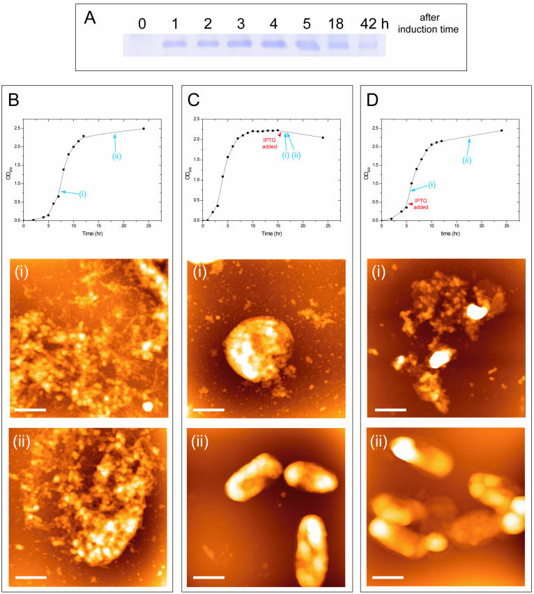
Complementation analysis of the Δdps nucleoid structures. W3110 (Δdps) cells carrying the plasmid pHM44 (Materials and Methods) were grown in LB medium and the expression of Dps protein was induced by IPTG (5 mM). (A) Immunoblot analysis of Dps protein. IPTG was added at OD = 0.2 (in the log phase) and the cells were collected at the indicated time after the induction and subjected to immunoblot analysis using anti-Dps antibody. (B–D) AFM analysis of the nucleoid structure. The expression of the Dps protein was not induced (B) or was induced with IPTG in the stationary phase (C) or in the log phase (D). The addition of IPTG did not affect cell growth (top panels). The cells were collected at the indicated time (i and ii) and subjected to observation by AFM (bottom panels). Scale bars, 1 µm.

