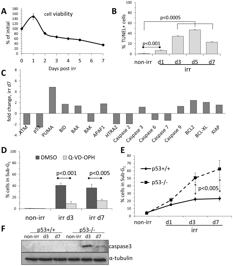
Figure 1. NSC undergo a delayed and p53-independent apoptosis upon irradiation.
(A)NSC cell density before (T0, normalized b as 100%) and after irradiation as measured by colorimetric MTT assay. Error bars: SD. (B)Apoptosis analysis by flow cytometrical TUNEL assay on non-irradiated (non-irr) NSCs and following irradiation. Error bars: SD. (C)Expression changes in apoptosis-relevant genes in NSCs at day 7 after irradiation compared to non-irradiated NSCs, based on microarray dataset originally published in [6]. (D)Flow cytometry analysis for apoptosis-associated DNA fragmentation (Sub-G1) of irradiated NSCs treated with pan-caspase inhibitor Q-VD-OPH (10 µM). Error bars: SD. (E)Flow cytometry analysis for apoptosis-associated DNA fragmentation (Sub-G1) of irradiated p53-deficient and isogenic wild type NSC. Error bars: SD. (F)Western blot analysis of irradiated p53-deficient and isogenic wild type NSC for apoptosis-associated appearance of active caspase 3. Loading control: α-tubulin.

