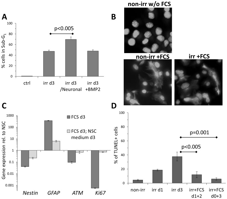
Figure 3. Apoptosis in irradiated NSC is dependent on culture conditions.
(A)Flow cytometry analysis for apoptosis-associated DNA fragmentation (Sub-G1) of irradiated NSCs, cultured in standard self-renewal medium, or supplemented with BMP2 (20 ng/ml), or switched to BDNF-containing Neuronal differentiation medium. Error bars: SD. (B)Representative immunofluorescence analysis of GFAP expression in non-irr and irr NSC subjected for 24 h to FCS. DNA was stained with DAPI. Magnification: 40×. (C)Gene expression analysis of astrocytes derived by 10% FCS from NSC (FCS d3), and when switched back to NSC culture medium, normalized against self-renewing NSC. Technical triplicates, error bars: SD. (D)TUNEL assay for irradiated NSC at day 3, cultured in unmodified (self-renewal promoting) medium or supplemented with FCS (astroglial differentiation medium), either immediately (d0+3) or 24 h after irr (d1+2). Error bars: SD.

