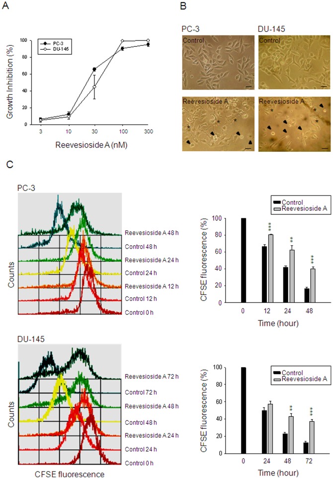
Figure 1. Effect of reevesioside A on cell proliferation.
Chemical structure of reevesioside A (A). The graded concentrations of reevesioside A were added to PC-3 and DU-145 cells for 48 hours (A) or a single concentration (50 nM) was added for 48 hours (B) or the indicated times (C). After the treatment, the cells were observed by microscopic examination (B) or the cells were fixed and stained for SRB assay (A) or labeled with CFSE for flow cytometric analysis. Data are expressed as mean±SEM of three to five determinations. ** P<0.01 and *** P<0.001 compared with the respective control. Arrowhead, cell apoptosis; star, cell differentiation; bar, 50 µm.

