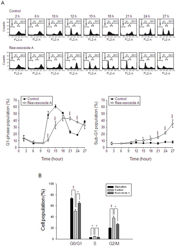
Figure 2. Effect of reevesioside A on cell-cycle progression.
(A) Synchronization of PC-3 cells was performed by thymidine block as described in the Materials and Methods section. Then, the cells were released in the absence (upper panel) or presence of 50 nM reevesioside A for the indicated times. Data are representative of five independent experiments. (B) DU-145 cells were incubated in serum-free medium for 48 hours (starvation) and then, 10% FBS was added in the absence or presence of reevesioside A for 18 hours. The cells were harvested for the detection of cell cycle population by flow cytometric analysis. Quantitative data are expressed as mean±SEM of five (A) or three (B) independent experiments. * P<0.05, ** P<0.01 and *** P<0.001 compared with the respective control.

