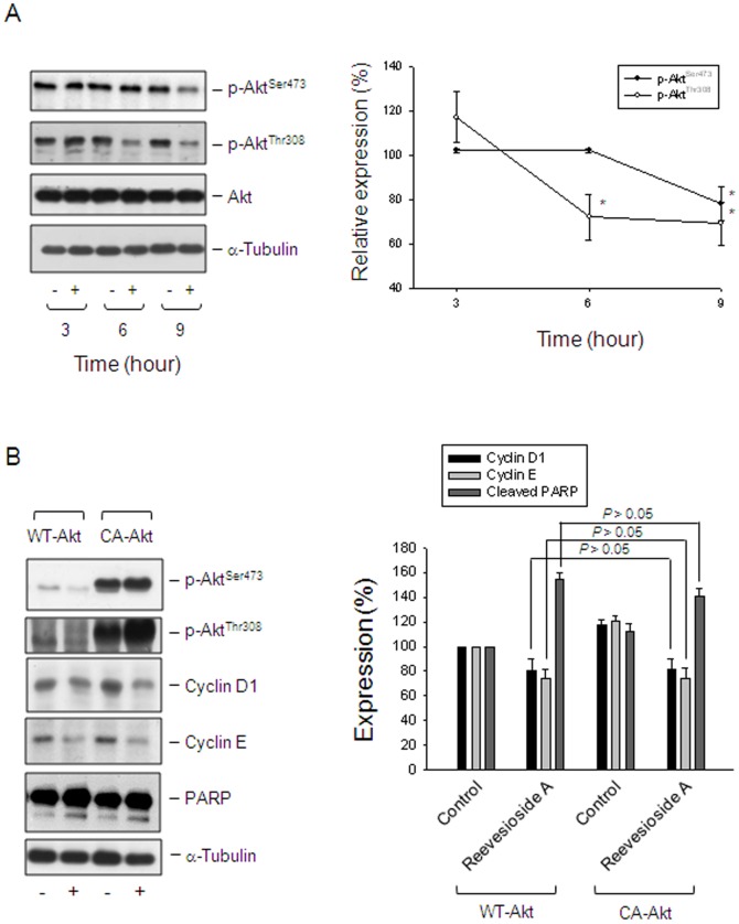
Figure 5. Determination of functional involvement of Akt.
(A) PC-3 cells were incubated in the absence or presence of reevesioside A (50 nM) for various times. The cells were harvested and lysed for the detection of the indicated protein by Western blot analysis. (B) PC-3 cells were transfected with the indicated plasmid. Then, the cells were treated without or with reevesioside A (50 nM) for 24 hours. After treatment, the cells were harvested and lysed for the detection of the indicated protein by Western blot analysis. The expression was quantified using the computerized image analysis system ImageQuant (Amersham Biosciences). The data are expressed as mean±SEM of three independent experiments. * P<0.05 and ** P<0.01 compared with 100% control. WT-Akt, wild type Akt; CA-Akt, constitutively active Akt.

