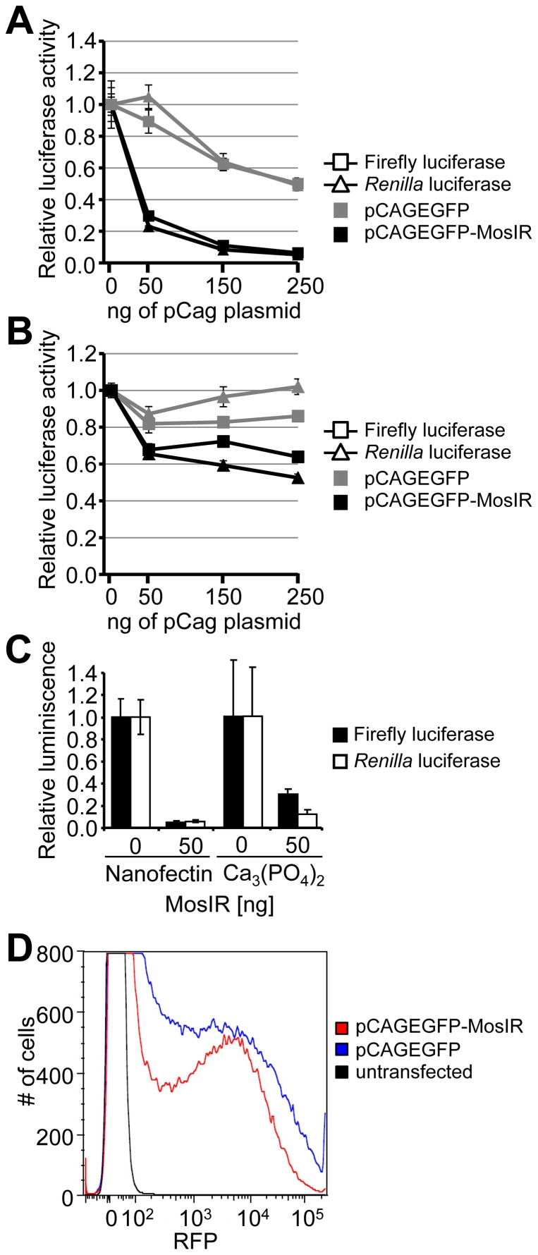
Figure 2. Suppression of co-transfected reporters in mammalian cells is general.
(A, B) Suppression of co-transfected reporters in HeLa cells (A) and mouse 3T3 cells (B). Cells were transiently transfected with a constant amount of firefly luciferase (square), Renilla luciferase (triangle) reporter plasmids, and increasing amounts of pCAGEGFP-MosIR or pCAGEGFP. Luciferase activities were measured 48 hours post-transfection. pBluescript was added to maintain a constant amount of transfected DNA. Both luciferase activities are shown relative to cells transfected with 0 ng of the pCAGEGFP-MosIR. Data are shown as an average of at least 3 experiments performed in triplicates. Error bars = SEM. (C) Suppression of co-transfected reporters is independent of the transfection method. HEK-293 cells were transiently transfected with a constant amount of firefly luciferase (black bars), Renilla luciferase (white bars) reporter plasmids, and 50 ng of pCAGEGFP-MosIR using Nanofectin or calcium phosphate transfection. Luciferase activities were measured 48 hours post-transfection. pBluescript was added to maintain a constant amount of transfected DNA. Both luciferase activities are shown relative to cells transfected with 0 ng of the pCAGEGFP-MosIR. Data show a typical experiment measured in triplicates. Error bars = SEM. (D) Suppression of a co-transfected RFP reporter. HEK-293 were transiently transfected with 150 ng of RFP reporter plasmid and 350 ng of pCAGEGFP or pCAGEGFP-MosIR. Shown is FACS analysis of RFP fluorescence 36 h post-transfection. The experiment was performed three times, a representative result is shown.

