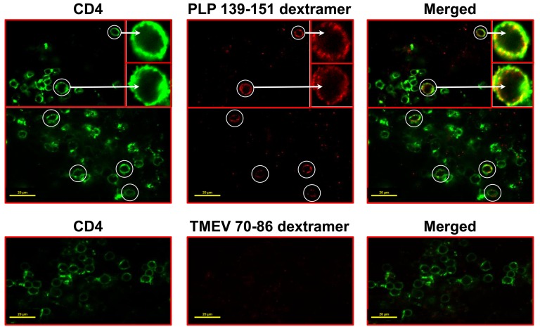
Figure 2. Detection of PLP-specific T cells by in situ staining with PLP 139–151 dextramers.
EAE was induced in SJL mice by immunizing the animals with PLP 139–151 in CFA. At termination, cerebrums collected from EAE mice were embedded in 4% agarose, and the sections were made using vibratome. After staining with cocktails containing either PLP 139–151 dextramers/anti-CD4 or TMEV 70–86 dextramers (control)/anti-CD4 and fixing with 4% PBS-buffered paraformaldehyde, sections were washed and mounted for examination by LSCM. Top panels: sections stained with PLP 139–151 dextramers and anti-CD4. Bottom panels: sections stained with TMEV 70–86 dextramers and anti-CD4. Left panel: CD4, green; Middle panel: dextramers, red; Right panel: merged (circles, dext+ CD4+ T cells; insets represent enlarged views of dext+ CD4+ T cells). Original magnification 1000×; bar = 20 µm.

