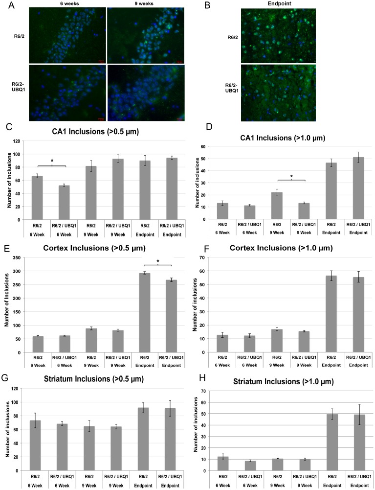
Figure 5. Ubiquilin-1 overexpression modifies aggregate load in the hippocampus and cortex but not the striatum.
(A) Representative fluorescence microscopy images of EM48 and DAPI stained cryostat sections of the CA1 region of the hippocampus in R6/2 transgenic and R6/2-UBQ1 double transgenic mouse at 6 weeks, 9 weeks and following end-stage euthanasia. Bar = 15 µm. (B) Similar to A, but showing representative sections from the dentate gyrus in end-stage mice. Bar = 15 µm. (C) Quantification of htt inclusions >0.5 µm in size in the CA1 region of the hippocampus at 6 weeks, 9 weeks and end-stage R6/2 and R6/2-UBQ1 double transgenic mice. The R6/2-UBQ1 double transgenic mice contained 22% fewer inclusions than R6/2 mice at 6 weeks (p = 0.04), but not at the other times. (D) Quantification of htt inclusions >1 µm in size in the CA1 region of the hippocampus at 6 weeks, 9 weeks and end-stage R6/2 and R6/2-UBQ1 double transgenic mice. The R6/2-UBQ1 double transgenic mice had 40% fewer inclusions at 9 weeks compared to R6/2 transgenic mice (p = 0.027). (E and F) Similar to B and C, but showing htt inclusions in the cortex. R6/2-UBQ1 double transgenic mice had 8.5% fewer inclusions greater than 0.5 µm at the end-stage of disease. (G, H) Similar to E and F, but comparing inclusions in the striatum. There was no difference in the number of inclusions in the striatum between the two genotypes at any time point.

