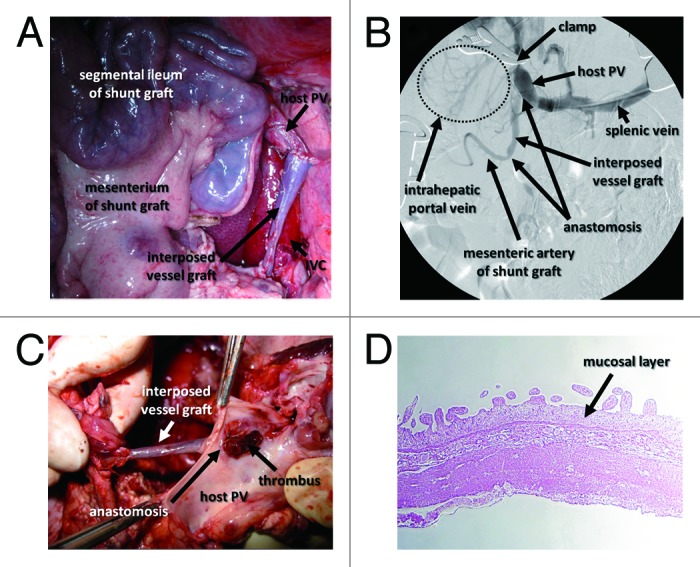
Figure 4. Intraoperative and postoperative findings of intestinal PCS in pigs. (A) Intraoperative image of the ileal graft and interposed vessel graft anastomosed to the host PV. (B) Intraoperative portography shows that temporary PH created by clamping the PV led to the visualization of the mesenteric artery of the intestinal graft. (C) The graft vessel interposed between the portal vein and small intestinal segment is occupied with a thrombus. (D) HE staining shows shortened villi and a thinned mucosal layer of the small intestinal segment.
