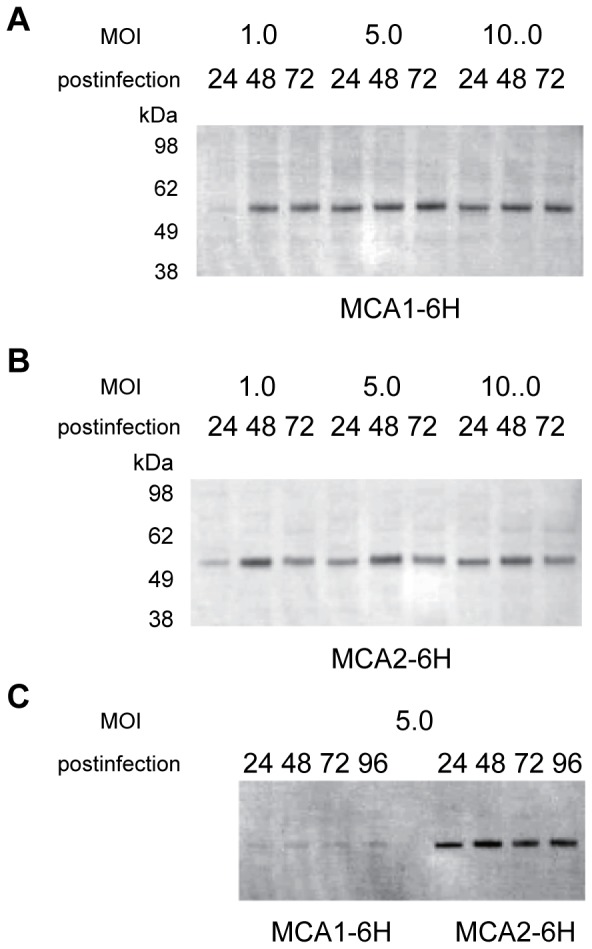
Figure 2. Expression profiles in Sf9 cells infected with a recombinant baculovirus.

Western blot analysis of the expression of the MCA1-6H (A, C) and MCA2-6H (B, C) proteins using anti-MCA1 (A), anti-MCA2 (B), and anti-6xHis tag (C) antibodies. Differences in the post-infection times and MOI are shown above the panels.
