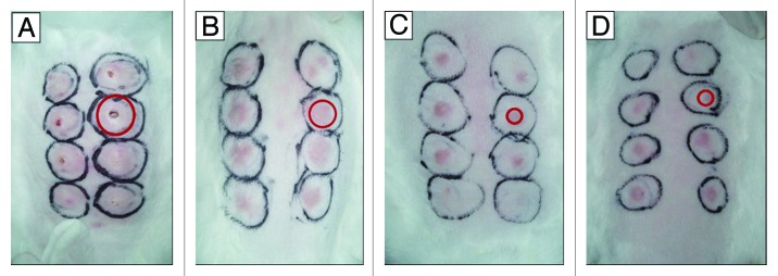
Figure 2. Representative photos of rabbits taken 21 d post intradermal challenges with Tp Nichols strain at eight locations on their backs. At week 10 after the first time of DNA vaccine immunization, 15 of the 18 rabbits in each group were challenged with Tp Nichols strain. Skin lesions were observed and measured at the challenged sites every 3 d. The red circle indicated one of the lesions in each panel. (A) Ulcerative and indurated lesions (a rabbit from the A1 group immunized with pcD); (B) Intermediate erythematous lesions (a rabbit from the B1group immunized with pcD/Gpd-IL-2); (C) The smallest, least indurated erythematous lesions(a rabbit from A2 group immunized with pGpd-IL-2); (D) The smallest, least indurated erythematous lesions(a rabbit from B2 group immunized with pGpd-IL-2).
