Figure 7.
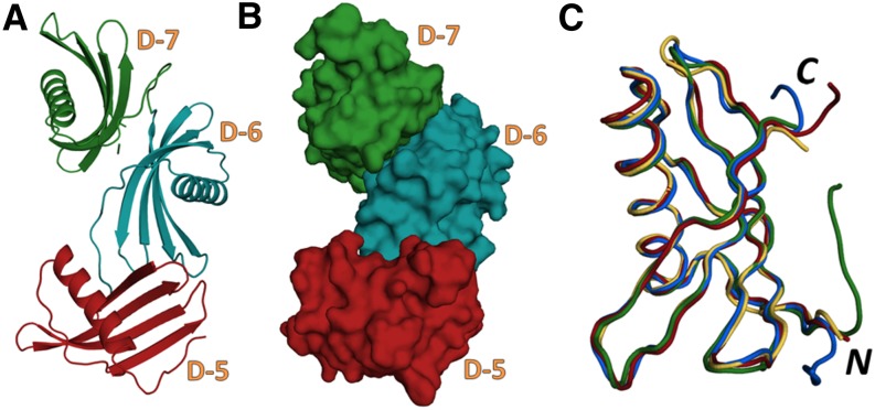
The Crystal Structure of PMC-567.
(A) and (B) Ribbon diagram (A) and space-filling model (B) representing the crystal structure of PMC-567. Domains 5, 6, and 7 are depicted as red, blue, and green, respectively.
(C) Backbone structures of PMC-5 (red), PMC-6 (blue), PMC-7 (green), and PMC-2 (orange) were superimposed. The N and C termini are labeled as N and C, respectively. These figures were generated using Open-Source PyMOL (v1.4).

