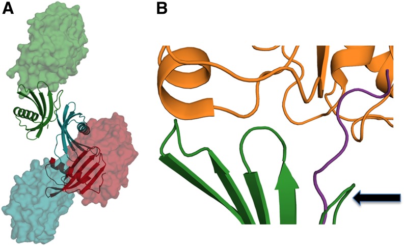
Figure 8.
Plausible Complex Formation between PMC-567 and Papain.
(A) Ribbon diagrams representing the core domain PMC-567 are colored by domain: 5 is red, 6 is blue, and 7 is green. Modeled papain molecules are represented as a space-filling model colored according to the PMC domain to which they are complexed. The positions of each papain were generated after superimposing the human stefin b in the papain complex structure (1STF) (Stubbs et al., 1990) with an individual PMC domain.
(B) Closer inspection of the molecular interface between PMC-7 (green) and papain (orange). The backbone of PMC-7 (green) has been superimposed with that of stefin b with the best molecular fitting. The N-terminal trunk of PMC-7 was curled around (arrow), which is significantly different to the extended conformation of stefin b (violet) observed in its papain complex (Protein Data Bank ID: 1SFT). These figures were generated using Open-Source PyMOL (v1.4).

