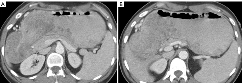
Figure 2.

CT images demonstrate extensive mucosal polyposis within the body and antrum of the stomach as well as portions of the fundus. (A) This CT image demonstrates the extensive gastric polyposis at the gastric outlet; (B) This CT image demonstrates the mucosal polyposis within the body, antrum, and portions of the gastric fundus.
