
Figure 1.
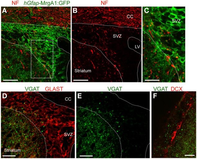
VGAT is expressed in the SVZ close to GFAP-positive cells and neuroblasts. (A,B) Confocal images of neurofilament immunostaining (NF, red, A) and GFP expression (green, A,B) in the SVZ in a coronal section from a hGfap-MrgA1:GFP mice. (C) Zoom from the white square shown in (A). (D,E) Confocal images of VGAT immunostaining (green, D,E) and GLAST (red, D), which decorates GFAP-expressing cells. GLAST is a glutamate transporter. (F) Confocal images of VGAT immunostaining (red) and DCX-expressing cells, neuroblasts (red). Scale bars: 40 μm (A,B), 20 μm (C,F), and 25 μm (D,E). CC, corpus callosum; LV, lateral ventricle.