
Figure 2.
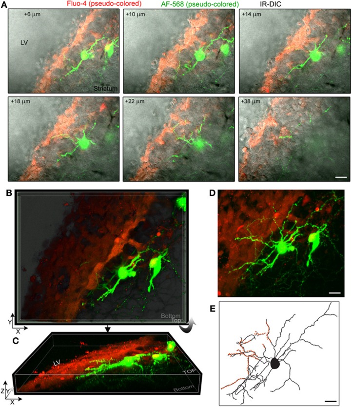
Striatal neurons project dendrites into the SVZ. (A) Images of single optical sections at different depths (indicated in the right corner) from the surface of the sagittal slice. Two striatal neurons were filled with Alexa Fluor-568 (pseudo-colored green) and the fluorescent dye Fluo-4AM (pseudo-colored red) was pressure applied in the SVZ. Not every SVZ cell is Fluo-4-positive. Images are overlaid on the IR-DIC. (B) 3D Z-projection using the blend mode in Imaris illustrating the 3D shape of the SVZ in a sagittal section. (C) 3D Y-projection of the image in B following a rotation up around the X axis. (D) Maximum intensity projection in Imaris. (E) Rreconstruction of one of the filled neuron with orange overlaid to marked the processes entering the SVZ. Scale: 30 μm (A,D,E).