
Figure 5.
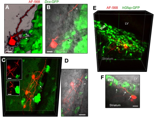
Striatal neurons send processes toward neuroblasts and GFAP-expressing cells. (A,B) Images of an AF-568-filled striatal neuron sending a process toward neuroblasts that are GFP-positive in Dcx-GFP mice. A represents a 3D projection in the blend mode of 21 sections (Z of 17 μm) and B represents 10 sections overlaid with IR-DIC. (C) Maximum intensity projection of three AF-568-labeled neurons in a coronal section from a Dcx-GFP mouse. Insets: zoom of the processes projecting toward neuroblasts (arrows in the main image) in one optical section. (D) Image of one optical section of the AF-568-labeled neuron and GFP fluorescence shown in (C) overlaid with IR-DIC image. The IR-DIC illustrates that neuronal processes also project toward GFP-negative cells. (E) Maximum intensity projection of a AF-568-labeled neurons in a sagittal section from a hGfap-GFP mice. The recorded striatal neuron sits below the SVZ and send a few processes into the SVZ. (F) Image of one optical section of the AF-568-labeled neuron and hGfap-GFP fluorescence shown in (E) overlaid with IR-DIC image. Arrows point to sites of contact between the neuronal processes and GFP-positive cell processes. Scale bars: 10 μm (A,B,C) and 15 μm (F).