Figure 2.

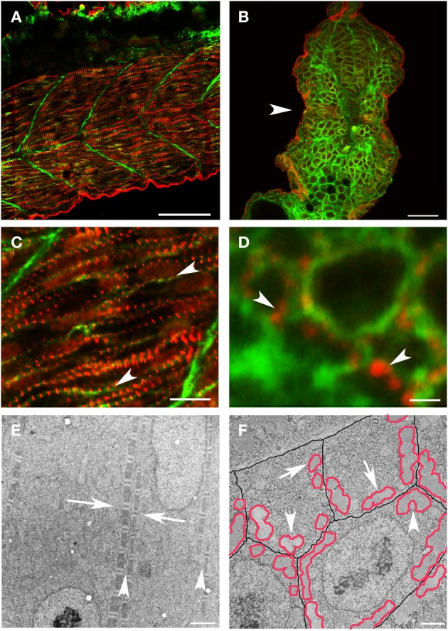
New myofibril assembly occurs adjacent to the sarcolemma in skeletal muscle. (A–D) Zebrafish embryos at 24 hpf were injected with lyn-GFP mRNA (which encodes for a GFP-fluorescent tagged protein that localizes to the cell membrane) and immunostained for α-actinin (red) and GFP (green) to mark the sarcomere and the sarcolemma respectively. Note that α-actinin dots (C,D: arrowheads) appear in a striated pattern immediately adjacent to the sarcolemma in both longitudinal optical (A,C) and transverse (B,D) sections. (E) Ultrastructural analysis reveals that there is a consistent relationship between new myofibrils that are forming on either side of the sarcolemma (E: arrowheads) and that there is a slight offset in the alignment of Z bands (E: arrows) across myocytes at this stage of development. In transverse sections (F), note that the myofibrils (outlined in red) form in close proximity to the sarcolemma (outlined in black) with preferential clustering in regions where three cells intersect (arrowheads). There are examples of myofibrils (arrows) that are not paired with a myofibril in an adjoining cell, suggesting that the organization of myofibrils across myocytes may not be direct and therefore may require patterning of the extracellular matrix to transmit architectural information. Scale bars are 50 μm (A), 20 μm (B), 10 μm (C), 1 μm (D), 2 μm (E), and 3 μm (F).
