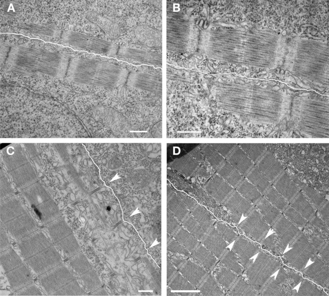
Figure 4.

Development of nascent costameres. Skeletal muscle from zebrafish embryos at 20 hpf (A,B) demonstrate a consistent offset in the alignment of the Z disks across adjacent myocytes (sarcolemma highlighted in white). Note that the distance from the myofibril to the membrane is consistent over the length of the myofibril. By 72 hpf (C,D), the membrane has a more undulated appearance and is closest to the myofibril overlying the Z disks (arrowheads) consistent with the maturation of costameric connections from the myofibril to the sarcolemma. Scale bars are 500 nm (A–C) and 2 μm (D).
