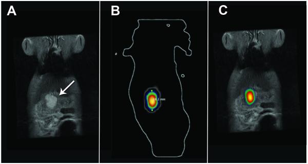
Figure 5.
Co-registration of 3T FSE T2-weighted MRI and 3D diffuse luminescence tomography (DLIT) images using 3D Multimodality Tools in Living Image Software. (A) Coronal FSE T2-weighted MR image of tumor (white arrow) and (B) Corresponding coronal 3D DLIT image; (C) Co-registered coronal FSE T2 MR and 3D DLIT image.

