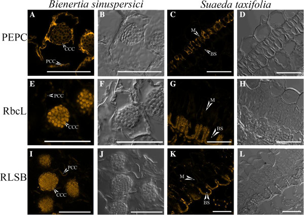
Figure 3.
Immunolocalization of C4 proteins in single cell type Bienertia and Kranz type Suaeda taxifolia. Confocal microscopy detection of the Alexa fluor 546 tagged secondary antibody, reacting to the primary antibodies indicated, showing the location of phosphoenolpyruvate carboxylase (PEPC, A-D), rubisco large subunit (rbcL, E-H), and rubisco large subunit mRNA binding protein (RLSB, I-L) in Bienertia(A, B, E, F, I, and J) and Suaeda taxifolia(C, D, G, H, K, and L). Images A, C, E, G, I, and K are Alexa Fluro 546 detection. Images B, D, F, H, J, and L are bright field view of sections. CCC = central compartment chloroplast, PCC = peripheral compartment chloroplast, M = Mesophyll Cell, BS = Bundle Sheath Cell. Scale bar = 50 μm.

