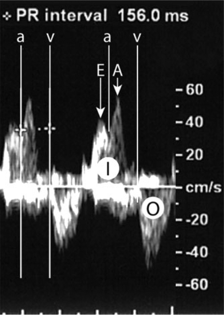
Figure 2.
Doppler MPRI. The left ventricular inflow (I) has an early flow wave (E) and a late flow wave during atrial contraction (A). The beginning of the mitral-valve A wave marks the beginning of the flow during atrial contraction (a). The left ventricular outflow (O) has a single wave and its beginning marks the beginning of the flow during ventricular contraction (v). The Doppler MPRI is measured from the beginning of the flow during atrial contraction (a) to the beginning of the flow during ventricular contraction (v)

