
Figure 3.
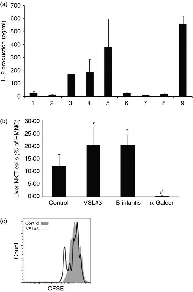
Probiotic antigens stimulate natural killer T (NKT) cells in vitro and in vivo. (a) Bacterial glycolipids extracted were co-cultured with NKT hybridoma. Interleukin-2 (IL-2) released by NKT hybridoma indicates their activation. 1 = medium only; 2 = artificial antigen-presenting cells (aAPCs) only; 3 = aAPCs loaded with low-dose VSL#3 extract; 4 = aAPCs loaded with high-dose VSL#3 extract; 5 = aAPCs loaded with high-dose VSL#3 extract plus α-GalCer; 6 = aAPCs loaded with low-dose Bifidobacterium infantis extract; 7 = aAPCs loaded with high-dose B. infantis extract; 8 = aAPCs loaded with high-dose B. infantis extract plus α-GalCer; and 9 = aAPCs loaded with α-GalCer. (b) Lipid extracts from VSL#3 or B. infantis, or α-GalCer (2 μg per mouse) were injected into C57BL/6 wild-type mice fed a normal diet. After 24 hr, the animals were killed and their hepatic NKT cells were evaluated as described in Fig 2. Means ± SD of the percentages of hepatic NKT cells (gated on CD3+ and CD1d Tetramer+) among hepatic mononuclear cells (HMNC) are shown (n = 5 per group). (c) A representative histogram of NKT cell proliferation assay. HMNC were labelled with CFSE and stimulated with aAPCs loaded with VSL#3 lipid extract or unloaded empty beads. *P < 0·025, #P < 0·002 versus control.