Figure 5.

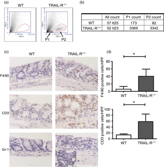
More extensive inflammatory cell infiltration in the colon tissues of tumour necrosis factor-related apoptosis-inducing ligand receptor deficient (TRAIL-R−/−) mice compared with wild-type (WT) mice after dextran sulphate sodium (DSS) administration. (a) Isolated colon lamina propria cells were analysed by flow cytometry. The cells from each group of mice were pooled from four individual mice. Target cells were gated based on forward light scatter (FSC) and side light scatter (SSC). Gate P1 represents lymphocytes, and Gate P2 represents mononuclear cells. (b) Counts of gated cells. (c) Immunohistochemical evaluation of colon tissue, where the brown colour represents F4/80-, CD3- and Gr-1-positive cells in WT and TRAIL-R−/− mice fed with 2·5% DSS for 5 days. For Gr-1 staining, inflamed lung tissue was used as a positive control. (d) Average numbers of F4/80+ cells (P = 0·033) and CD3+ cells (P = 0·030) in colonic tissue sections. Data represent the mean ± SEM of four mice per group, and three high-power fields (HPF) were counted per mouse.
