
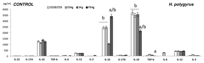
Figure 3. Concentration of IL-2, IL-6, IL-22, IL-17A, IL-10, IL-12 and TGF-β in cultures of MLN cells from uninfected and day 15 infected with H. polygyrus mice. Cells (5 × 10⁵ ⁄ well) were treated with excretory–secretory (ESAg), somatic antigen (SAg) and fraction 9 (F9) and cultured for 7 d. Concentrations of cytokines were measured by specific ELISA. A representative result from three independent experiments is shown. Bars represent the mean ± SE of five mice of representative experiment (n = 5). Statistical significance between groups (control and infected) was assessed by Anova. ap < 0.05 compared with untreated cells (MEDIUM) within the same group; bp < 0.05 compared with cells with other group treated by the same manner.