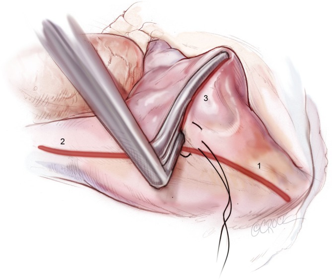
Figure 4.

Ablations of the [1] IVC, [2] SVC and [3] right atrial free wall directed towards the AV groove near the acute margin of the heart. Ablations are performed using a bipolar RF clamp through a purse-string suture placed midway between the SVC and IVC. IVC, inferior vena cava; SVC, superior vena cava; AV, atrioventricular; RF, radiofrequency.
