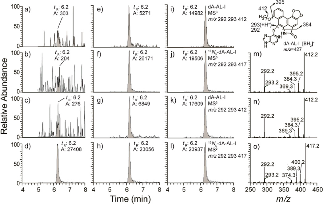
Figure 4.
Representative mass chromatograms at the MS3 scan stage and product ion spectra of dA-AL-I in freshly frozen and FFPE human kidney DNA (5 µg) spiked with 15N5-labeled internal standard at a level of 5 adducts in 108 bases (d, f, h, j, and l). The chromatograms were reconstructed from the extracted ions of dA-AL-I (MS3 at m/z 292, 293, 412) and [15N5]-dA-AL-I (MS3 at m/z 292, 293, 417). Three untreated calf thymus DNA samples served as the negative controls (a–c); freshly frozen human kidney tissue (e, i) and FFPE kidney tissue (g, k) from 2 human patients with UUC. Product ion spectra were acquired at MS3 scan stage for dA-AL-I in frozen (m) and FFPE tissue (n), and for the internal standard [15N5]-dA-AL-I (o). The structure of the aglycone [BH2]+ adduct of dA-AL-I and proposed mechanisms of fragmentation of the adduct at the MS3 scan stage are shown in the top right corner.

