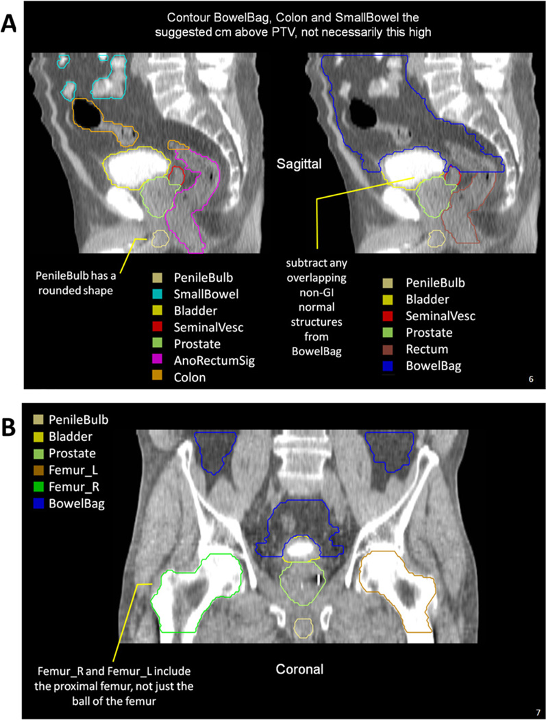
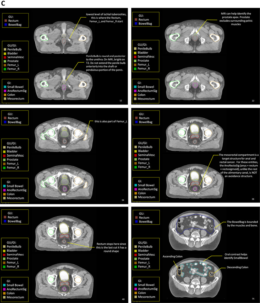
Fig. 1.
Highlights from the Radiation Therapy Oncology Group (RTOG) male pelvis normal tissue atlas: sagittal view (A), coronal view (B), and axial views (C). The full atlas is available on the RTOG Web site. GI = gastrointestinal; GU = genitourinary; MRI = magnetic resonance imaging; PTV = planning target volume.


