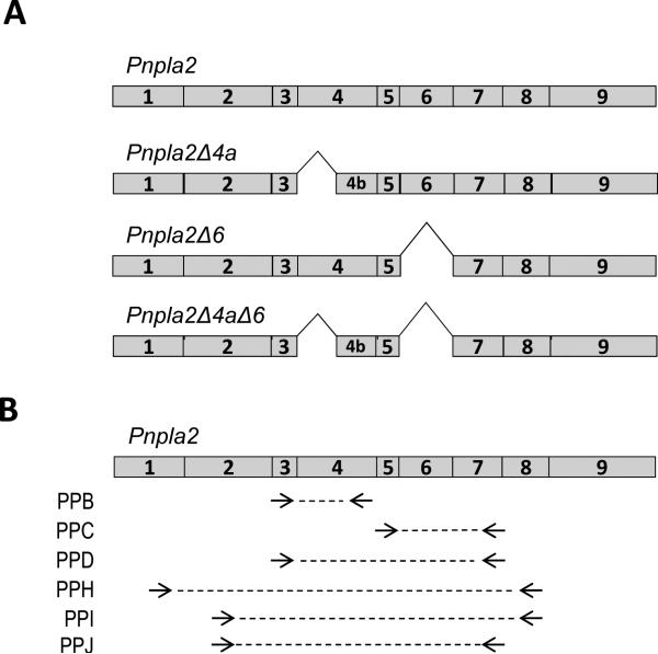
Figure 1.
Predicted Pnpla2 splice variants and primer pair locations. A) Scheme of full-length mouse Pnpla2 and the splice variants predicted by EST databases. Shown are Pnpla2 transcripts with exon 4a (E4a) deletion (Pnpla2Δ4a), with exon 6 (E6) deletion (Pnpla2Δ6) and with both E4a and E6 deletion (Pnpla2Δ4aΔ6). B) Arrow heads correspond to the 3’ end of primer pairs (PP) B, C, D, H, I, and J on the full-length mouse Pnpla2 transcript. PPB and D have the same forward primer; PPI and J have the same forward primer; PPC, D, and J have the same reverse primer; and PPH and PPI have the same reverse primer. The dotted lines illustrate the amplified DNA fragment. The numbers inside the boxes correspond to the exon number. See Table 1 for primer sequences.

