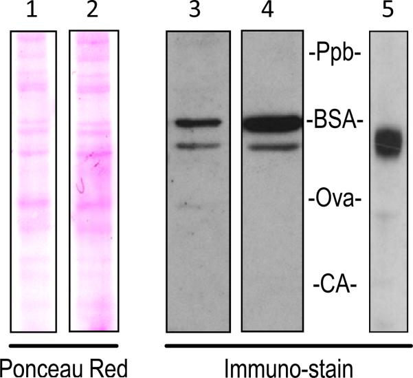
Figure 6.
PEDF-R protein in mouse 661W cells. Equal amounts of 661W total cell lysate were run on the same 4-12% polyacrylamide Bis-Tris gel. Nitrocellulose membranes were either stained with Ponceau Red total protein stain (Lane 1) and then probed with AF5365 anti-PEDF-R (Lane 3) or stained with Ponceau Red (Lane 2) and then probed with SAB2500132 anti-PEDF-R (Lane 4). Recombinant human PEDF-R was also run on a 4-12% Bis-Tris gel and probed with anti-Xpress (Lane 5). Labels of Ppb (phosphorylase b), BSA (bovine serum albumin), Ova (ovalbumin), and CA (carbonic anhydrase) show the migration pattern of the prestained molecular weight markers.

