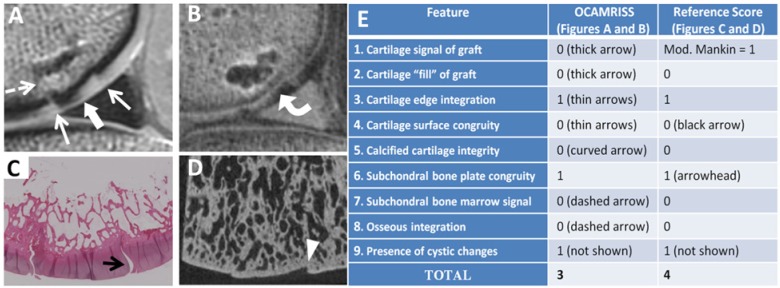
Figure 1.

Medial femoral condyle allograft with fresh storage and good performance (OCAMRISS TS9-MRI 3 points and TS9-REF 4 points; cartilage stiffness = 4.2 MPa). Sagittal proton density (PD)–weighted image (A), sagittal 3D ultrashort echo time (UTE) subtraction image (B), hematoxylin and eosin stain (C), and micro–computed tomography (D) demonstrate features as listed in the accompanying table (E).
