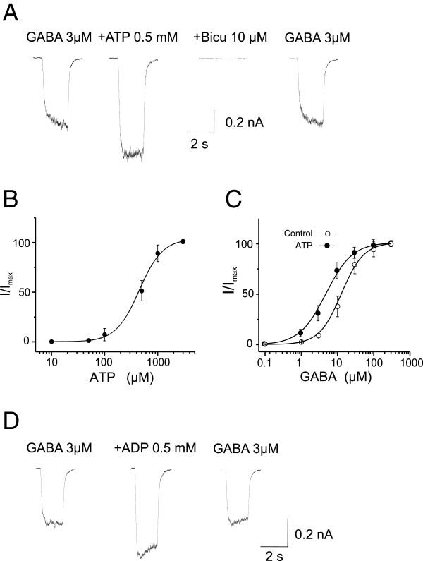
Figure 6.
Extracellular ATP and ADP potentiates function of recombinant GABAARs. Whole-cell voltage-clamp recordings were performed in HEK293 cells transiently expressing rat recombinant α1β2γ2 GABAARs at a holding membrane potential of -60 mV. GABA currents were induced by fast perfusion of GABA at various concentrations. A, Representative current traces showing the potentiation of GABA (3 μM) currents by co-perfusion of ATP (0.5 mM). The increased current was blocked in the presence of bicuculline (10 μM) and returned to the control level after ATP washout. B, The dose-response curve showing the dose-dependent potentiation of the GABA (3 μM) currents by various concentration of ATP. All values were normalized to the maximal GABA currents (Imax) induced at 3 mM of ATP. Each data point is the mean ± SEM of normalized GABA currents at the indicated ATP concentrations (n = 6). The solid line is the best fit of the data to the Hill equation. The EC50 and H were 390 ± 48 μM and 1.66 ± 0.1, respectively. C, ATP potentiation of GABAAR activity is GABA dose-dependent. GABA dose-response curves were constructed in the absence (Control) and presence (ATP) of ATP (0.5 mM) from five cells. ATP shifted the curve to the left and reduced EC50 from 11.6 ± 3.3 μM to 5.8 ± 2.1 (P < 0.01) without change in H (control: 1.57 ± 0.1; ATP: 1.62 ± 0.1; P > 0.05). All values were normalized to the Imax induced by GABA (300 μM). D, ADP mimics ATP, potentiating the activity of recombinant GABAARs. Representative current traces showed that application of ADP (0.5 mM) reversibly increased currents induced by GABA (3 μM). On average, it increased the currents by 147.5 ± 15.9% (P < 0.01; n = 5).

