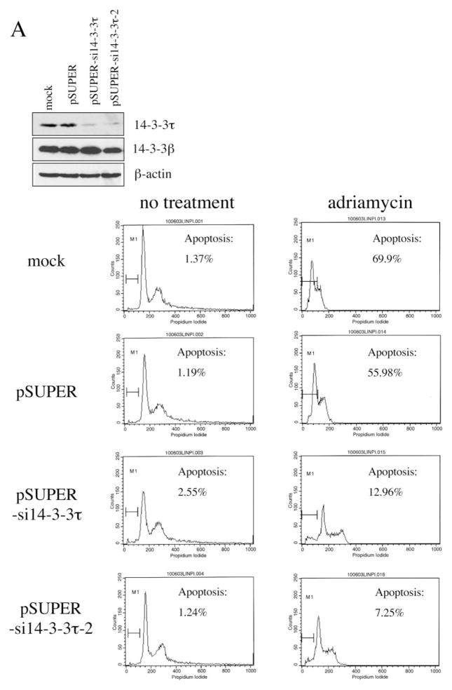
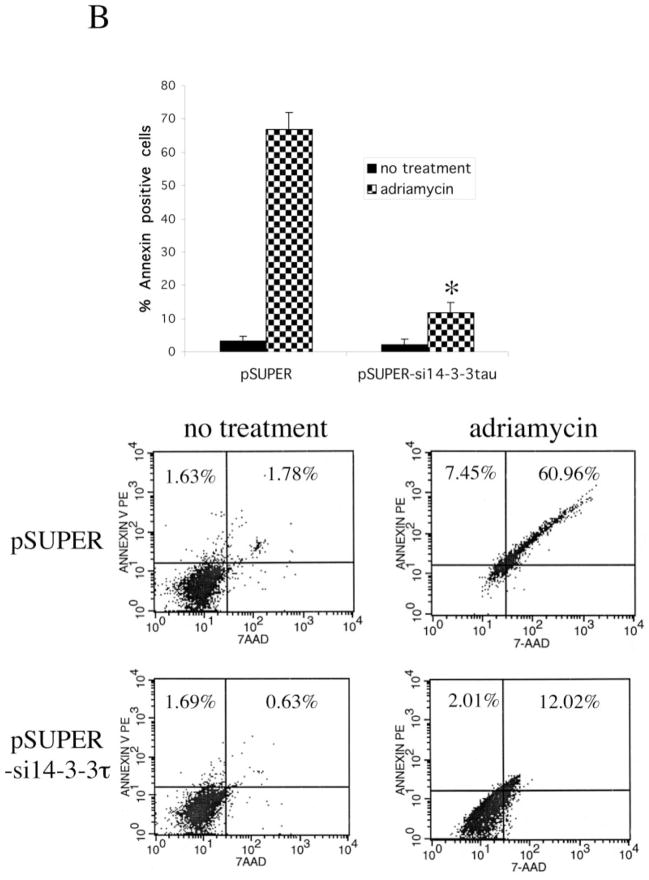
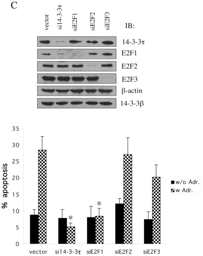
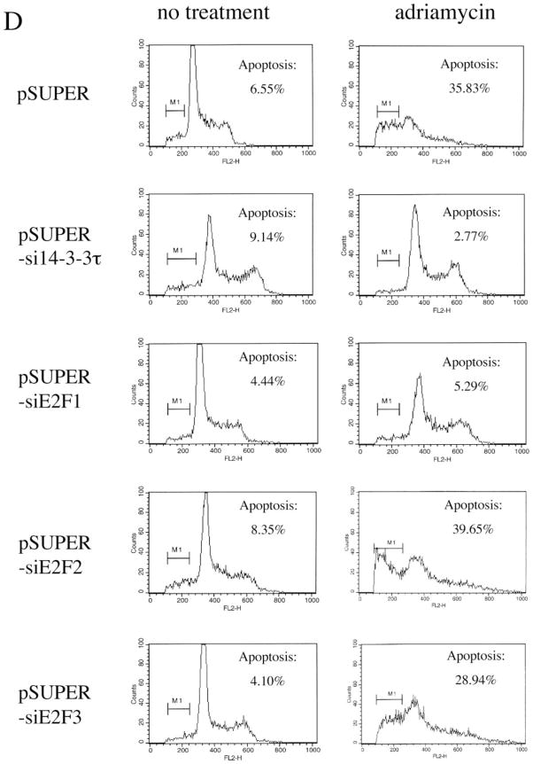
Fig. 6. A role for 14-3-3τ in adriamycin (Adr)-induced apoptosis.
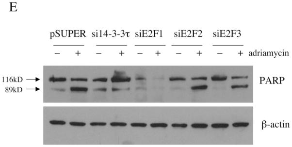
A, HEK293 cells were mock-transfected or transfected with pSUPER or pSUPER vectors expressing two different 14-3-3τ siRNAs and treated with adriamycin (5 μM) for 48 h. Apoptotic cells were counted by propidium iodide-flow cytometry. Inhibition of 14-3-3τ expression but not 14-3-3β expression in this experiment was confirmed by Western blot analysis (top). B, HEK293 cells were transfected with pSUPER or pSUPER-si14-3-3τ and pcDNA3-GFP, and treated with adriamycin as in A. Cells were then stained with annexin V-PE and 7-AAD. GFP-positive cells were gated and analyzed by flow cytometry. Top, the data shown are the mean ± S.E. of three independent experiments. *, p < 0.05 (t test) compared with the pSUPER vector group. Bottom, a representative flow cytometry profile. C, HEK293 cells were transfected with the plasmids expressing 14-3-3τ, E2F1, E2F2, or E2F3 siRNA and treated with adriamycin (5 μM) for 24 h. Apoptosis was scored by propidium iodide-flow cytometry. The data shown are the mean ± S.E. of three independent experiments. *, p < 0.05 (t test) compared with pSUPER vector group. Knockdown of 14-3-3τ or E2F by corresponding siRNA in these experiments was confirmed by immunoblotting (top). D, a representative propidium iodide-flow cytometry profile from the experiments described in C. E, the cell lysates from the experiments described in C were subject to Western blot analysis for PARP cleavage. β-actin immunoblot was performed to confirm equal protein loading.

