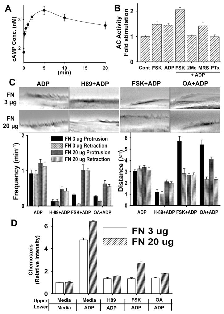
Figure 2.
(A) ADP transiently stimulates Adenylyl Cyclase (AC) activity. BV2 cells were stimulated with 100 μM ADP for the time indicated and then cAMP concentration was measured with CatchPoint Cyclic-AMP fluorescent Assay kit. (B) ADP-induces AC activation through the Gi/o-coupled P2Y12 receptor. BV2 cells that were pretreated with inhibitors and cells were then lysed and subjected to cAMP assay. (C) FSK, OA, and H-89 have an inhibitory effect on ADP-induced membrane ruffling. Cells were pretreated with 10 μM FSK, 1 μM Okadaic acid (OA) or 30 μM H-89 for 20 min and then stimulated with 100 μM ADP. Lamella dynamics was then analyzed by kymographs. (D) FSK, OA, and H-89 have an inhibitory effect on ADP-induced chemotaxis of microglia cells. Transwell chamber membranes were coated with 3 μg/ml or 20 μg/ml fibronectin for 8 hr. Cells were then assayed for migration toward 100 μM ADP in the continued presence of H-89, FSK and OA. Averages of five independent experiments are shown.

