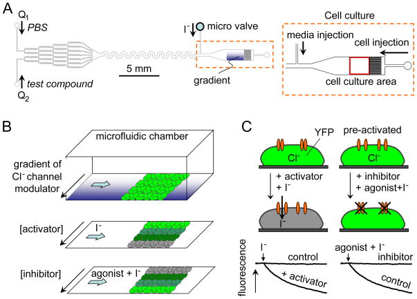
Figure 1.
Cell-based microfluidics fluorescence assay of chloride channel activity. A. Microfluidic chamber design showing gradient-generating channels at the left and cell culture area at the right. B. Assay method showing perfusion with a concentration gradient of chloride channel modulator followed by rapid exchange (top) to generate an inwardly directed iodide gradient of activator (middle) or inhibitor (bottom). C. Schematic showing fluorescence quenching of a cytoplasmic YFP iodide sensor following addition of iodide with an activator (left) and inhibitor (right).

