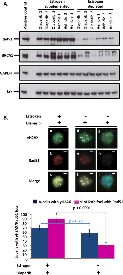
Figure 5. Decreased levels and function of Rad51 may drive the sensitivity of PTEN-null tumors to PARP inhibition in estrogen depleted conditions.
(A) Western blot reveals diminished expression of both Rad51 and BRCA1 in lysates of tumors from estrogen deprived compared to estrogen supplemented mice regardless of treatment with Olaparib or vehicle. 3T3 and HeLa lysates were positive controls for Rad51 and BRCA1 respectively. Erk was used as a loading control. Expression of GAPDH was not altered by estrogen levels, suggesting that reduced proliferation with estrogen deprivation is not responsible for changes in Rad51 or BRCA1 levels. (B) Rad51 was efficiently recruited to Olaparib-induced sites of DNA damage (marked by γH2AX foci) in PTEN-null endometrial epithelial cells supplemented with estrogen (a–c). In estrogen deprived cells, low (d–f) or no (g–i) levels of Rad51 incorporation into γH2AX foci was observed compared to estrogen supplemented mice (p<0.0001). Olaparib induced γH2AX foci formation was unaffected by estrogen deprivation (p=0.29). Scale bars equal 10 μm.

