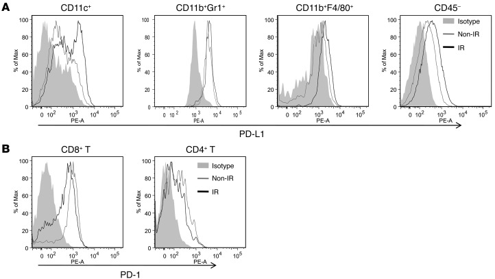
Figure 1. The profile of PD-L1 and PD-1 expression in tumor microenvironments is altered after IR.
BALB/c mice were injected s.c. into the flank with 1 × 10⁶ TUBO cells. On day 14, mice were locally treated with one 12-Gy dose of IR. Three days after IR, tumors were removed and digested into single-cell suspensions, which were blocked with anti-FcR mAbs and then subjected to surface staining. PD-L1 expression on myeloid cells and tumor cells (A) and PD-1 expression on T cells (B). Representative data are shown from three (A and B) experiments conducted using 3 mice per group.

