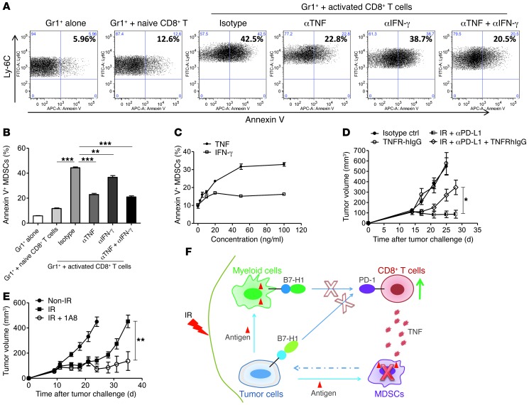
Figure 6. CD8+ T cells induce the apoptosis of MDSCs through TNF-α following combination therapy.
(A and B) Isolated CD8+ T cells derived from naive BALB/c mice were stimulated with anti-CD3 and anti-CD28 for 6 hours. Then, Gr1+ cells purified from the spleen of TUBO-bearing mice were added for an additional 21 hours’ coincubation. Representative dot plots (A) and the percentage (B) of annexin V+ in MDSCs are shown. Cells were gated on a CD11b+Ly-6C+ cell population. **P < 0.01; ***P < 0.001. (C) Percentage of annexin V+ in MDSCs after treatment with increasing concentrations of TNF. Gr1+ cells purified from the spleen of TUBO-bearing mice were treated with different concentrations of TNF or IFN-γ for 21 hours. (D) The inhibition of tumor growth with combination therapy was abrogated after the treatment of TNFR-hIgG. TUBO tumor–bearing mice were treated with IR and antibodies as described in Figure 2A. 500 μg TNFR-hIgG (etanercept) was administered every 4 days for a total of three times starting on the first day of IR. *P < 0.05. (E) Depletion of MDSCs augmented the efficacy of IR treatment. C57BL/6 mice were injected s.c. on day 0 with 1 × 10⁶ MC38 cells. Tumors received 20 Gy on day 9, and 300 μg of depletion antibody against MDSCs (clone 1A8) was administered every 2 days for a total of four times starting 1 day prior to IR. **P < 0.01. Representative data are shown from two (A–E) experiments conducted with 4 (D) or 5 (E) mice per group. (F) Schematic of proposed mechanism for tumor destruction induced by IR and PD-L1 blockade.

