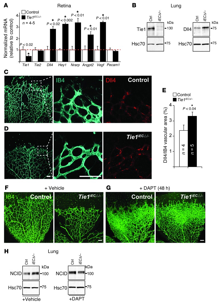
Figure 7. Tie1-deficient retinal phenotype involving upregulation of Dll4/Notch is abolished by Notch inhibitor.
(A) Quantitative RT-PCR analysis of control and Tie1iECΔ/– (Cdh5(PAC)-cre/ERT2 and Pdgfb-icre/ERT2 deletors) retinas at P6. mRNA levels were normalized to Cdh5 to compensate for the decreased vascular area in the Tie1iECΔ/– retinas and expressed relative to control levels (assigned as 1; red dashed line). (B) Representative Western blot analysis of lungs (see Supplemental Figure 19 for quantification). n = 5–8 (control); 6–14 (Tie1iECΔ/–). (C and D) IB4 (cyan) and Dll4 (red) staining of (C) control and (D) Tie1iECΔ/– angiogenic retinal fronts. (E) Quantification of Dll4-positive endothelium (expressed as a percentage). (F and G) IB4 (green) staining of retinas from control and Tie1iECΔ/– (Pdgfb-icre/ERT2 deletor) P6 pups treated with (F) vehicle or (G) DAPT for 48 hours. (H) Representative Western blotting for Notch1 cleaved intracellular domain (NCID) from vehicle- and DAPT-treated lungs. n = 3–4 lungs/genotype. Scale bars: 50 μm. Error bars denote SD (A) or SEM (E). Significant differences are shown by asterisks, with P values indicated.

